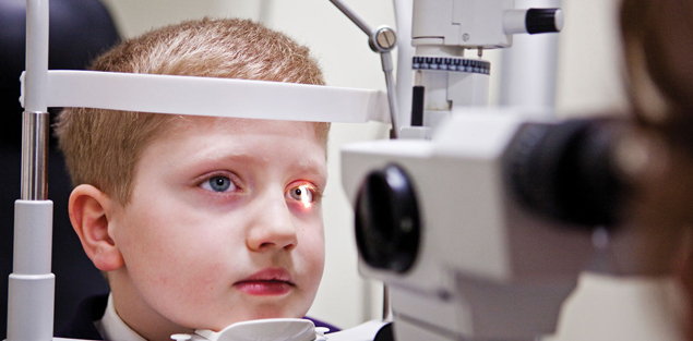
- Cataract
- Cataract in Children
- Protecting your Child’s Eyesight
- Diabetes
- Diabetic Retinopathy
- Microphaco
- Multifocal and Foldable Lens Implants
- Lasik
- Squint Evaluation and Surgery
- Cornea Services
- Oculoplastics
- Botox
- AMD
- Glaucoma
- Low Vision AID
- Contact Lenses
Cataract Surgery

Sight is our most precious gift, enabling us to enjoy the beauty of the world in which we live. For most people with poor vision from cataract, the prospects of regaining good vision and resuming normal daily activities are excellent.
Symptoms:
- Frequent changes in Eyeglass prescription
- Poor Night Vision
- Needing a brighter light to read
- Double Vision in one Eye
- Fading Colours
How is a Cataract Detected?
A thorough eye examination by an ophthalmologist detects the presence and the extent of a cataract. Other conditions that could additionally compromise vision can also be detected, particularly problems involving the cornea, retina or the optic nerve.
How fast does a Cataract Develop?
Cataract development varies among individuals and may even vary between the two eyes. Most cataracts associated with the aging process develop over years. Cataracts in younger patients and in those with diabetes may develop rapidly.
Treatment of a Cataract?
Surgery is the only way a cataract can be removed. No dietary supplements, medications, exercises or optical devices have been proven to prevent or cure cataract.
When should Surgery be done?
Surgery should be considered when the cataract causes enough visual disturbances to interfere with daily activities, such as job requirements, safe driving and household activities. Based on these needs and examination findings, the patient and the ophthalmologist should decide together when surgery is appropriate.
It is not true that cataracts need to be ‘ripe’ before they can be removed. In fact advances in the field of cataract surgery have made it easier to remove an immature cataract than a mature [‘ripe’] cataract.
What should I expect from a Cataract Surgery?
Cataract surgery is a microscopic surgery usually performed under local or topical anaesthesia. The cloudy lens is removed by Phacoemulsification, which is a micro-incision technique of cataract surgery wherein a probe breaks the cataract into tiny pieces and sucks them out. If a foldable implant is inserted the incision size is smaller (2.8 – 3.0 mm) as compared to an approximately 5 mm incision to accommodate a non-foldable lens. Both incisions are self-sealing and need no stitches. We will help you decide as to which lens should suit you best.
What are the benefits phacoemulsification Surgery for Cataract?
The wound is smaller [resulting in faster healing and visual rehabilitation], the surgical time is reduced, and no stitches are required. There is minimal or no post-operative discomfort and a quick return to your normal routine.
Cataract Surgery can now be customized to reduce and usually eliminate dependence on glasses for distance, intermediate vision and reading Plan your Cataract Surgery at Isight Eyecare and Surgery:
I nitial Assessment
S election of Lens
I nvestigations
G o for the actual Procedure
H ave regular post op follow up
T otal recovery
Cataract in Children

Cataract is defined as an opacity in the crystalline lens or its capsule, which in turn causes a drop in vision.
While majority of cataracts occur in the middle aged or elderly, pediatric cataracts are also seen quite frequently. The incidence of congenital cataract is approximately 1 in 5000 births.
Congenital (present at birth) cataracts occur when the lens didn’t form properly. Acquired cataracts are caused by abnormal interactions among the proteins that make up the lens, which, over time, cause clumping, specks, opacities and/or cloudy areas to form.
About 25 percent of the time, congenital cataracts have a genetic cause, and may accompany a metabolic, hormonal or chromosomal abnormality (e.g., Down syndrome). Another 25 percent of the time, cataracts are hereditary, which means that the child’s mom or dad also had a cataract in childhood.
Often cataracts are idiopathic – meaning they occur for no identifiable reason.
While each child may experience symptoms differently, things to look out for are reduced vision, frequent falls or clumsiness, disinterest in visual activities like reading or watching TV, or in younger babies there could be an involuntary eye movement or squinting.
If your child is too young to complain about vision problems, you might notice a white pupil if you shine a flashlight into her eye.
Most babies are looking around and tracking things with their eyes nicely by the time they’re 3 or 4 months old. If you get the sense that your baby isn’t seeing and responding appropriately, that may be a tip off that something is wrong. Jiggling eye movements may also be a sign of a cataract or other eye problem.
Strabismus, or misaligned eyes (eyes that are not looking in the same direction), usually aren’t a sign of cataracts – most often, they’re just the result of a newborn’s poor coordination. However, sometimes strabismus occurs if there is a cataract in just one eye.
These cases are picked up by an alert parent who notices that the child cannot see very well or that there is a preferential gaze which the child adopts always. Sometimes they are picked up on routine screening in school or a regular visit for refraction.
There are three important parts in the management of the child with a cataract: Preoperative evaluation, surgery, and visual rehabilitation.
A complete preoperative evaluation of both eyes is required. Some cataracts are unilateral and the health of the apparently normal eye requires careful evaluation as well. An eye with a cataract may have other associated abnormalities which require assessment preoperatively in order to provide the most reasonable expectation for visual outcome. An ultrasound (sound waves), used to image the inside structure of the eyes, is needed in some cases.
Surgery is the mainstay of treatment. The treatment is cataract extraction with intra ocular lens implantation. This may be combined with vitrectomy in some cases.
Problems?
Since the child eye is still growing the estimation of the exact lens power to be implanted is difficult to determine. The lens implantation in children requires a compromise that accounts for the age of the child and the target post operative refraction. However these children will become more myopic over the years and eventually may need a secondary procedure sometime in future to correct the resultant refractive error.
Unlike adults, cataract surgery in children requires general anesthesia. The post operative care involves wearing spectacles and also patching the eye in some cases.
The deal with congenital cataract is that the approach to each case is tailor made to suit that patient best.
Regular follow up is essential to the successful recovery of a patient.
Despite adequate optical rehabilitation and patching therapy, misalignment of the eyes (strabismus) often develops. This may require eye muscle surgery to improve the alignment.Periodic evaluations are required to detect any other post-operative complications (for example, secondary cataract, inflammation, glaucoma, retinal detachment). A child who has undergone cataract surgery requires close monitoring of visual development and will need changes in glasses or contact lenses as the eye matures. The parents must accept the responsibility for the optical rehabilitation, patching therapy, and continued follow-up care that is required to maximize the visual development in the operated eye. Without the participation of parents, optimal vision will not be realized.
Good visual outcome after surgery depends on many factors, including patients age, type of cataract and timing of surgery. A strong post operative visual rehabilitation program to prevent amblyopia.
Amblyopia Management:
This should begin immediately in the post operative period. Corrective glasses or contact lenses and patching of the better eye is needed and decided on an case to case basis.
Overall, the visual outcome for children with cataracts is much more optimistic now than it was a few years ago. However, some eyes will have associated abnormalities, which limit the visual potential that can be obtained, despite early surgery and adequate visual rehabilitation.
Protecting your Child’s Eyesight
Sports and your child’s eye – If your child is involved in contact sports or ball games, eye protection is a must. Polycarbonate Plastic for lenses and special safety materials for frames is recommended.
Swimming and your child’s eye – Chlorine in pools can cause irritation. Infections can be contracted in freshwater. Avoid these harmful effects by using water tight swimming goggles.
VDUs (Visual Display Units) and your child’s eye – Include computer and television screens. Prolonged work with these may give rise to eyestrain, headaches, burning, itching, blurring of vision, double vision, nausea and fatigue. Here are a few recommendations:
For Computers
- Check position to minimize reflection and Glare
- Preferred viewing distance is 60 to 80 cms
- Place the screen at eye level or slightly lower
- Take periodic breaks, 5 to 10 minutes every hour
For Televisions
- Do not watch TV in a dark room
- Preferred viewing distance is 3 to 4 meters
Lighting and your child’s eye
While reading, the light source should be positioned behind your child. At a desk, preferably use a shielded light since this prevents light from shining directly in the eyes. The light should not be glaring. The reading material should be at a distance of 12-14 inches.
Preserving your child’s eyesight
Loss of vision is the most feared disability, particularly when someone in the family has an eye problem or a ‘high number’. However, many of the problems leading to visual loss are related to lifestyle choices. Vision depends on a clear lens, rich microcirculation to the retina, and visual nerve cells of the retina called rods and cones. These are adversely affected on exposure to toxins and oxygen free radicals, and poor dietary habits.
Common eye problems in children
- Refractive errors (spectacle number) account for 80% of visual impairment in children. This includes Myopia or near-sightedness, where distance vision is blurred, but a child can usually see well for reading or other near tasks. This occurs most often in school age children. The prescription for glasses will indicate a minus sign before the prescription (for example, – 2.00).
Another problem is Hyperopia (far-sightedness), where problems such as crossing of the eyes, blurred vision, or discomfort may develop. However, most children are far-sighted early in life and it becomes a concern only in excessive cases where the focusing muscles are not able to keep the vision clear. A prescription for hyperopia will be preceded by a plus sign (+3.00).
Astigmatism is also common and is caused by a difference in the surface curve of the eye. Instead of being shaped like a perfect sphere (like a basketball), the eye is shaped with a greater curve in one axis (like a rugby ball) and this causes fine details to look blurred or distorted. Here, the prescribed glasses have greater strength in one direction of the lens than in the opposite direction.
Anisometropia causes children to have a different prescription in each eye. This can create a condition called lazy eye, where the vision in one eye does not develop normally. Glasses (and sometimes patching) are needed to ensure that each eye can see clearly.
- Lazy eye or Amblyopia is seen in 1% of children. It is a failure of the eye to connect to the brain due to lack of use of one eye during infancy and childhood. Therefore, the vision in the amblyopic eye remains poorer than in the normal eye. To solve this, the poorer seeing eye is forced to be used by patching the preferred eye and combined with the use of glasses where necessary. Amblyopia must be corrected by 9 years of age by which time it becomes permanent. To detect and treat ambylopia, it is necessary to examine pre-school age children.
- Squint or Strabismus, also called crossed-eyes, means that the eyes are not aligned but are pointing in different directions. They may either point inwards towards the nose (esotropia), outwards to the ear (exotropia) or up or down (vertical strabismus). The problem can be constant or intermittent. However, intermittent strabismus occurs in infants during the first few months of life, especially when the infant is tired, since they are still learning to focus their eyes and to move them in a coordinated fashion.
Most babies outgrow this intermittent strabismus by the age of 3 months. For other cases, various treatment options are available, including exercises for the eyes, called orthoptics. Some of the ncommon eye problems in children include cataract, glaucoma and retinal problems, which are treated through different methods.
Start with the diet
All children need the same nutrients, no matter what their age. Give your child a healthy diet with an emphasis on fresh vegetables and fruits, whole grains, beans, seeds, and nuts. The nutrients that they provide are important for overall as well as eye health. For non-vegetarians, add eggs and a small amount of fish. Avoid those addictive sweets along with the sodas, cookies, pies, ice cream, and candy. Junk food is a prescription for disaster when it comes to eyesight. Staying with a mostly vegetarian diet of natural, unprocessed foods will help preserve the circulation to the retina, the rods and cones, and the lens.
Add Exercise for Circulation
Regular exercise helps to maintain a low blood pressure and preserves the blood vessels, which the retina is rich in. It also improves circulation to the heart through the coronary arteries. Exercise also helps to support the health of the cells that line the arteries, called endothelium. These cells help relax the blood vessel muscles and maintain blood flow.
Protection from ultraviolet light
Eyes undergo damage to the lens and the retina on exposure to ultraviolet light. Protecting the eyes from too much sun exposure may help delay any degeneration of the lens and the retina. However, some sun exposure is healthy as long as it is in moderation. Sunlight is important for the skin for the production of vitamin D. A wide-brimmed hat and UVfiltering sunglasses (in older children) provide protection for the eyes.
Supplements for the eyes
Over 25% of childhood blindness in India is due to Vitamin A deficiency. Many dietary supplements improve vision and help protect the macula (the area of the retina with the sharpest vision) and the lens such as vitamins E and C, and the trace minerals selenium and zinc. Others are the amino acid taurine, carotenoids such as beta-carotene, lycopene (found in tomatoes), lutein (from spinach), and the anthocyanosides (related to bioflavonoids) found in grapes.
Recommendations for Eye examination in children
A pediatrician should examine a new born’s eyes.An ophthalmologist should be asked to examine all premature infants.Vision screening should be done between 3 and 3½ years of age.Detailed eye examination must be done if there is appearance of symptoms of visual impairment.
This leaflet is intended to provide general information for patients.
Diabetic

As part of an eye examination, your doctor may include a diagnostic procedure called fluorescein angiography to identify leaking blood vessels.
In fluorescein angiography, your doctor injects a dye into a vein in your arm. The dye circulates through your eyes, making the blood vessels in your retina easy to identify. Your doctor can pinpoint areas where normal blood vessels have become closed or have broken down and are leaking fluid. A camera with special filters takes flash pictures every few seconds for several minutes, providing your doctor with useful images.
Your doctor also may request an optical coherence tomography (OCT) examination. This noninvasive imaging scan provides high resolution images of the retina that show, for example, the thickness of the retina and whether fluid has leaked into retinal tissue. OCT exams can be useful both as a diagnostic tool and as a way of monitoring treatment effectiveness.
Diabetic Retinopathy

Diabetes affects your body from head to toes. This includes your eyes. The most common and most serious eye complication of diabetes is diabetic retinopathy, which may result in poor vision or even blindness.
“Retinopathy” is the medical term for damage to the tiny blood vessels (capillaries) that nourish the retina, the tissue at the back of your eye that captures light and relays information to your brain. These blood vessels are often affected by the high blood sugar levels associated with diabetes.
Nearly half of people with known diabetes have some degree of diabetic retinopathy. The longer you have diabetes, the more likely it is you’ll develop diabetic retinopathy. Initially, most people with diabetic retinopathy experience only mild vision problems. But the condition can worsen and threaten your vision.
The threat of blindness is scary. But with early detection and treatment, the risk of severe vision loss from diabetic retinopathy is small. You can take steps to protect your sight if you have diabetes. These include a yearly eye examination and steps to keep your blood sugar, blood pressure and blood cholesterol under the best possible control.
Diabetic retinopathy is caused by damage of the blood vessels in the retina. Nonproliferative diabetic retinopathy develops first when the vessels in the eye become larger in certain spots. Proliferative diabetic retinopathy is the advanced form of the disease where new blood vessels start to grow in the eye.
Diabetic Retinopathy is caused due to diabetes problems that inadvertently affect the eye at a certain stage. Diabetes eye problem that requires treatment if some or more of the symptoms occur: blurry or double vision, dark or blank spots, pain or pressure in eyes and trouble seeing from the corner of the eyes.
Diabetic Retinopathy – Treatment
If you have mild nonproliferative diabetic retinopathy, you may not require treatment right away. However, your eye doctor will want to closely monitor your retina. Proliferative diabetic retinopathy requires prompt surgical treatment.
Photocoagulation
The goal of photocoagulation, also known as laser treatment, is to stop the leakage of blood and fluid in the retina and thus slow the progression of diabetic retinopathy and vision loss. The decision to use the procedure depends on the type of diabetic retinopathy you have, its severity and how well it may respond to treatment.
Your doctor may recommend photocoagulation if you have:
- Diabetic macular edema, a swelling that involves or threatens the center of the retina.
- Severe nonproliferative diabetic retinopathy, especially if you have type 2 diabetes.
- Proliferative diabetic retinopathy.
- Neovascular glaucoma.
In photocoagulation, a high-energy laser beam creates small burns in areas of the retina with abnormal blood vessels to help seal any leaks. The procedure takes place in your doctor’s office or in an outpatient surgical center. Before surgery your eye doctor dilates your pupil and applies anesthetic drops to numb your eye. In some cases he or she numbs your eye more completely by injecting anesthetic around and behind your eye.
First, your chin and forehead are rested in an examination device called a slit lamp. This is a microscope that uses an intense line of light (slit) to allow your doctor to clearly view portions of your eye. Then, your doctor places a medical contact lens on your cornea — the layer of clear tissue at the front of your eye — to help focus laser light onto the sections of the retina to be treated. Fluorescein angiographic photographs may serve as maps to show where the laser burns should be placed. During the procedure you may see bright flashes from the short bursts of high-energy light.
To treat macular edema, the laser is focused on spots where blood vessels are leaking near the macula. The doctor makes “spot welds” to stop the leakage. If the leaks are small, the laser is applied directly to specific points where the leaks occur (focal laser treatment). If the leakage is widespread or diffuse, laser burns are applied in a grid pattern over a broad area (grid laser treatment).
Shortly after laser treatment, you can usually return home, but you won’t be able to drive, so make sure to arrange for a ride. Your vision will be blurry for about a day. Even when laser surgery is successful in sealing the leaks, new areas of leakage may appear later. For this reason you’ll have follow-up visits and, if necessary, additional laser treatments.
Immediately following laser surgery to treat macular edema, small spots caused by the laser burns may appear in your visual field. The spots generally fade and disappear with time. If you had blurred vision from macular edema before surgery, you may not recover completely normal vision.
Panretinal Photocoagulation
For proliferative diabetic retinopathy, doctors use a form of laser surgery called panretinal or scatter photocoagulation. With this technique the entire retina except the macula is treated with scattered laser burns. The treatment causes the abnormal new blood vessels to shrink and disappear. Thus it reduces the chances of a vitreous hemorrhage and traction retinal detachment. Panretinal photocoagulation is usually done in two or more sessions.
You may notice some loss of peripheral vision afterward. Panretinal photocoagulation is a trade-off. Some of your side vision is sacrificed to save as much of your central vision as possible. You may also notice difficulties with your night vision.
Vitrectomy
A vitreous hemorrhage may clear up on its own. But if the hemorrhage is massive and doesn’t clear, a vitrectomy may help to restore your sight and may allow the application of needed laser treatment.
In this procedure your surgeon uses delicate instruments to remove the blood-filled vitreous. A vitreous cutter cuts the tissue and removes it, piece by piece, from your eye. The tissue that is removed is replaced with a balanced salt solution to maintain the normal shape and pressure of the eye. A light probe illuminates the inside of the eye. The surgeon performs the procedure while looking through a microscope suspended over the eye. In this way the vitreous blood is removed to re-establish clear vision.
A vitrectomy is also used to remove scar tissue when it begins to pull the retina away from the wall of the eye. This allows a detached retina to settle back and flatten out. Your eye doctor may decide not to operate on a retina detached by scar tissue if the detachment is located away from the macula and doesn’t appear to be progressing.
During a vitrectomy the surgeon may also use a laser probe to perform panretinal photocoagulation. This can help prevent renewed growth of abnormal blood vessels, bleeding and scar tissue formation.
Vitrectomy can be performed under local or general anesthesia. Sometimes it is necessary to inject a bubble of expandable gas into the eye cavity. As the gas bubble expands, it pushes on the retina and helps it reattach. You may be required to remain in a face-down position for several days until the gas bubble spontaneously goes away. Your eye will be red, swollen and sensitive to light for some time after surgery. For a short time afterward, you’ll need to wear an eye patch and apply medicated eyedrops to help the healing. Full recovery may take weeks.
Microphaco

A revolutionary leap in cataract management – it allows cataract surgery via a 2.2 mm incision only.
This technology is relatively new but there are many scientific studies to prove its efficiency and it is known to have outstanding results. This advanced technology is now available at ISight Eyecare and Surgery for the benefit of our cataract patients.
The advantages are a quicker recovery and less possibility of post operative requirement of prescription glasses.
Talk to our counsellors for more information on this fabulous procedure.
Through Micro Phaco Technology a very small incision is require to remove Cataract from Eye. The tip of the instrument is introduced into the eye through ultra small incision. Localized high frequency waves are generated through this tip to break the cataract into very minute fragments and pieces, which are then sucked out through the same tip in a controlled manner. A thin ‘capsule’ or shell is left behind after cleaning up of the entire opaque cataract. After removal of Cataract, Surgeon inserts Inra Ocular Lens (IOL) in patient’s eye.
Type of Intra Ocular Lenses (IOL) for Cataract Surgery
- Foldable IOL Micro Phaco Surgery
- Aspheric Foldable IOL Micro Phaco Surgery
- Toric Foldable IOL Micro Phaco Surgery
- Multi-Focal Foldable IOL Micro Phaco Surgery
Multifocal and Foldable Lens Implants

With the advent of new technology and gadgets, our lifestyles have also undergone a change.
A typical day would involve an hour at the gym, followed by a busy day at work, brainstorming with colleagues, making power point presentations, checking important mails on the smartphone.At the end of a work day, check your emails in the car, go the club for a round of golf or a swim, head home and crash out.Well, as you can easily tell – our parents and grand parents were not leading such hectic or fast paced lifestyles.
With globalisation, our dependency on gadgets and smartphones has really gone up. So also our shopping happens in a supermarket or mall, with rows of stuff stacked on shelves.
Our leisure activities have changed too. This kind of a lifestyle needs not just a good lens implant but a great one – which will ideally cater to the distance, near and also intermediate range of vision.
The good news is that there are options now available in the form of multifocal lens implants . Please visit the doctor for more details on this topic.
Lasik Surgery

One of the most popular ways to correct vision is with a procedure called LASIK (laser in-situ keratomileusis), which uses a laser to change the curvature of the cornea (outer window of the eye). LASIK has quickly become the procedure of choice for most patients because they recover quickly and have fewer side effects and complications than with other methods of vision correction. In fact, most LASIK patients notice a significant improvement in their vision soon after surgery. LASIK removes tissue within the cornea to treat low to high levels of nearsightedness, farsightedness and astigmatism.
LASIK is an acronym for Laser-Assisted In Situ Keratomileusis, a refractive surgery procedure performed by Ophthalmologists intended for correcting the vision in order to reduce a person’s dependency on corrective devices such as glasses or contact lenses. The procedure is usually a preferred alternative to PRK, or photo refractive keratectomy, as it requires less time for full recovery, and the patient experiences less pain overall.
The first step in the procedure consists of mapping the corneal surface with a computer controlled scanning device to determine the exact shape. Then the appropriate amount of tissue which needs to be removed is calculated, such that upon replacing the flap there is no need (or less need) for corrective devices like spectacles and lenses.
LASIK is a procedure that permanently changes the shape of the cornea, the clear covering of the front of the eye, using an excimer laser. A blade, called a microkeratome, is used to cut a flap in the cornea. A hinge is left at one end of this flap. The flap is folded back revealing the stroma, the middle section of the cornea. Pulses from a computer-controlled laser vaporize a portion of the stroma and the flap is replaced. When the cornea is properly reshaped, it will then better focus light onto the retina. This results in improved vision
Squint

What is Squint (Strabismus)?
Squint is a misalignment of the two eyes so that both the eyes are not looking in the same direction. This misalignment may be constant, being present throughout the day, or it may appear sometimes and the rest of the time the eyes may be straight.
It is a common condition among children. It may also occur in adults.
What causes Squint?
The exact cause of squint is not really known. The movement of each eye is controlled by six muscles. Each of these muscle acts along with its counterpart in the other eye to keep both the eyes aligned properly. A loss of coordination between the muscles of the two eyes leads to misalignment. This misalignment may be the same in all directions of gaze, or in some conditions the misalignment may be more in one direction of gaze, e.g., in squint due to nerve palsy.
Sometimes a refractive error hypermetropia (long sight) may lead to inward deviation of the eye. Poor vision in an eye because of some other eye disease like cataract, etc. may also cause the eye to deviate. Therefore it is important in all the cases of squint, especially in children, to have a thorough eye checkup to rule out any other cause of loss of vision.
What is Binocular Vision?
Under normal circumstances, when both the eyes have good vision and they are aligned properly, they focus on the same object. Each of the eyes sends picture of the same object, viewed from a slightly different angle. These two images reach the brain, where they are fused to form a single three-dimensional picture with depth perception. This is known as binocular single vision.
What are the problems with Squint?
When the eyes are not aligned properly, each of the eyes is focusing on a different object and sends signal to the brain. These two different images reaching the brain lead to confusion and may have either of the two effects:
A child would ignore the image coming from the deviated eye, and thus sees only one image. But in the process, he loses the depth perception. This suppression of the image from the deviating eye results in poor development of vision in this eye, which is known as amblyopia.
An adult can not ignore the image from either eye, and therefore has double vision. This can be very annoying and may interfere with work.
What are the symptoms of Squint?
In a child, the parents may notice the deviation of eyes. It is important to remember that the eyes of a newborn are rarely aligned at birth. Most establish alignment at 3-4 weeks of age. Therefore squint in any child who is more than one month old must be taken seriously and should be evaluated by an ophthalmologist.
Adults may notice double vision, or misalignment of the eyes.
How is Squint Diagnosed?
The squint is diagnosed by the ophthalmologist. He or she would do a few special tests to confirm the squint, to try and find out the cause and to quantify the amount of deviation. In some cases there may be a false appearance of squint due to broad nasal bridge in a child. An ophthalmologist will be able to differentiate between a true squint and false squint.
What is the treatment for Squint?
- Preserve or restore vision
- Straighten the eyes
- Restore binocular vision
First of all, the eyes are checked to see if they have any refractive error that may be responsible for squint. If there is any significant refractive error present, it is treated first. In some cases (accommodative squint) a correction of refractive error is all that may be required to treat squint.
Next the eyes are checked for presence of amblyopia. It is important to treat the amblyopia before the surgery for squint. The parents are explained about the importance of this treatment, as their cooperation is very crucial for the success of this treatment.
The squint is treated by surgery of either one or both the eyes. The surgery involves weakening or strengthening of the relevant muscles to restore the balance and to get a good coordination. In some cases with double vision, prisms may be added in the glasses to ease the symptoms.
Cornea Services

We provide a range of cornea services at our clinic.
What are the common ailments of the cornea for which I need to see a cornea specialist?
- Corneal Foreign Body
- Pterygium
- Dermoid
- Dry Eyes
- Keratoconus
- Allergic Conjunctivitis
- Vernal Kerato Conjunctivitis
- Herpes Simplex
- Herpes Zoster
- Contact Lens Allergy
- Corneal Ulcer
- Corneal Edema
What are the special equipments I will be tested on?
- Pachymeter
- Keratometer
- Slit Lamp
- Fluorescein Staining
- Esthesiometer
- Topography – Orbscan
Oculoplastics

Oculoplastics, or oculoplastic surgery, includes a wide variety of surgical procedures that deal with the orbit (eye socket), eyelids, tear ducts, and the face.
When do I need to visit an oculopastic surgeon?
- Watery Eyes
- Loss of Eyelashes
- Mis directed Eyelashes
- Droopy Eyes – ptosis
- Bulging Eyes – proptosis
- Lid twitching – blepharospasm
- Wrinkles around the corners of the eyes – crows feet
- Baggy or Puffy Eyes
- Chronic sticky discharge from the Eyes- Dacryocystitis
- Inability to close the eyelids post facial paralysis – bells palsy – lagophthalmos
- Tumours or cysts on the eyelids or conjunctiva
- Thyroid Patients
Can I get Botox done around the eyes?
Yes. Botox is a safe procedure for wrinkles around the eye area and for lid twitches.
It is also used in some cases of squint and diplopia.
Botox
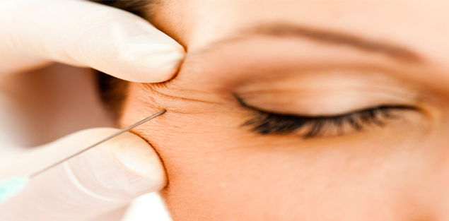
Can I get Botox done around the eyes?
Yes. Botox is a safe procedure for wrinkles around the eye area and for lid twitches.
It is also used in some cases of squint and diplopia.
AMD

ARMD:
What is ARMD?
AGE RELATED MACULAR DEGENARATION IS A disease that affects the macula in elderly persons. It is a fairly common condition and is a leading cause for irreversible vision loss in developed nations.
There are two types, dry AMD and wet AMD.
Can it affect me?
It can affect anyone, after the age of 60years.
It is equally common in men and women.
Potential risk factrs are systemic disesases like hypertension and diabetes; as well as high myopes.
What are the symptoms?
Well, essentially you will experience a drop in vision or a fixed black spot in the central visual field.
You may see someones face such that you cant see the nose clearly but can see the ears well, and similarly for other objects in your visual field.
There is sometimes a sudden appearance of a large black spot or a blindspot that can be quite intimidating for the patients.
Often , prior to this happening, you may complain of seeing wavy or distorted images instead of clear ones with sharp lines.
How do I know I have ARMD?
If have these symptoms you must visit an eye specialist right away. You will be tested on an amslers grid as a screening procedure.
You will also undergo a dilated retinal evaluation and may need specific tests like a macular OCT.
Whats the PROGNOSIS?
The outcome is quite variable. Rarely does the condition improve. Stalling the progression of disease is the goal of treatment here.
With newer treatment options the success rates are fairly high.
Dry type, unfortunately, cannot be treated . it is best managed by providing special glasses as low visual aids.
The wet type can be treated with intravitreal injections.
These injections halt the progression of disease by preventing new vessel formation and reducing the cnvm- choroidal neovascular membranes. These injections need to be repeated at monthly intervals till the desired effect is achieved. The results are judged with serial OCT scans done at monthly visits.
Alternately, lasers and surgery are done in severe and non responsive cases.
Glaucoma

DON’T LET GLAUCOMA DARKEN YOUR LIFE
Glaucoma is a condition in which eye pressure rises and causes damage to your optic nerve and visual field. Most people with glaucoma have no symptoms or pain from this increased pressure.It is important to see your eye doctor regularly so that glaucoma can be diagnosed and treated before long-term visual loss occurs. If detected early, the damage can be limited.
How is Glaucoma Treated?
Glaucoma treatment may include prescription eye drops, laser procedure, or microsurgery of the eye.
What is Glaucoma locally known as?
Various terminologies like “Jhamar”,”KaalaPaani”,”KaalaMotiya”,”Eye Pressure”.
Will I become blind?
Visual loss once set in cannot be reversed.With correct treatment lowering of IOP and regular follow up ,further damage is stalled or delayed. It is very rare for a patient to become blind if there is a regular follow up and good compliance with treatment schedule.
Am I at risk?
Yes if you :-
• Have a family history of glaucoma
• use spectacles.
• Have diabetes
• Takes steroid medications/asthma pumps
• Are over age 40
What diet should I follow?
1. Consume abundant amounts of fruits and vegetables.
2. Consider eating fish or nuts rich in omega-3 PFA, which appear to reduce risk.
3. Avoid drinking large amounts of liquid in a single take. It is preferable to drink small amounts in the course of the day.
4. Consume moderate amounts of red wine, black chocolate and green tea.
5. Avoid coffee and caffeinated beverages or colas if you already have glaucoma.
How do I know if I have glaucoma?
Watchout for these symptoms
• Seeing halos around lights
• Redness in the eye
• Eye that looks hazy (particularly in infants)
• Pain in the eye
• Narrowing of vision (tunnel vision)
Should I get a screening test done anyways?
Yes, it is advisable for all to get a glaucoma work up done, at least once, so that it is picked up in the earliest stages, before any damage sets in.
This test is called OCT.
How Is Glaucoma Diagnosed?
After routine eye examination test like:
• Intra ocular pressure (IOP) measurement
• Pachymetry (corneal thickness assessment)
• Perimetry (visual field testing)
• Optic disc analysis (digital retinal scan or OCT test)
Low Vision AID
coming soon.
Contact Lenses
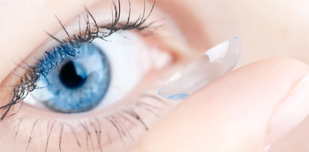
Contact lenses are available in a wide range of powers and materials to suit all types of individual needs.
If you wish to wear lenses for cosmetic reasons or for even improving your quality of vision, they are a great option.
1.When do I need lenses?
Once you are mature enough to handle the lenses and take care of them well, you can start using them.many school kids start using lenses by the age of 14-15 years onwards.
2.What are the benefits over glasses ?
Well, the field of vision is larger with lenses. When you wear glasses, the peripheral field is lost due to the edge effect of the glass rim. This is restored with lenses.
3. Will lenses hurt?
No. Initially you may experience minimal discomfort just like you would with the new pair of shoes. Over time and with a good fit, lenses are extremely comfortable.
4.Can I sleep with lenses on?
No, never. Even a short nap is highly discouraged.
5.What about taking care of lenses?
Our experts will guide you in day to day care of lenses and advise you on which solution works best for you.
6.How do I decide, Daily/Monthly/weekly- Its very confusing. How do I know which to get?
Stress not here are some useful guidelines for you to decide:-
1. If you plan to wear your lenses everyday, for most of the day, a monthly modality works well.
2. In addition to this, If You work in mall excesses screen time you would much rather opt for a 15 days Modality.
3. If you plan to wear them occasionally, a daily lense is the best. A daily lense, does not gather any debris and so, is most suited for infrequent wear .
7.I have a cylindrical Number, so can I still wear lenses ?
Yes, of course, Toric contact lenses will help you to see better.
8.What if I have keratoconus, which ttype of lens should I Opt for?
Customized rigid lenses will work best for you.
9.I am over 40 and I wear bifocal glasses. So, can I wear Contact Lenses ?
Yes, you can still wear lenses. Monovision contact lenses will help to solve your problem.
10.Can I wear lenses after Lasik Surgery?
Yes, occasionally you can wear colored lenses.